Leftover Halloween Candy and Pumpkins: What to Do Next
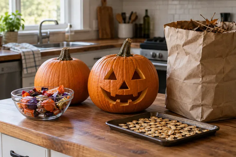
Sort leftover Halloween candy safely, share or repurpose sealed treats, and choose food use or compost for pumpkins based on their condition.
By combining MERFISH imaging with expanded microscopy, scientists have opened up a new way to study bacteria at the single-cell level. This new achievement allows for more in-depth observations of how bacteria activate different types of genes in response to their environment, providing insights into bacterial behavior, antibiotic resistance, and infection strategies.
How Bacteria Work
How do bacteria – whether the beneficial ones that inhabit our bodies or the dangerous strains that cause disease – coordinate their activities? A new study has provided new insights by combining advanced genomic microscopy with a groundbreaking technique to track the genes that bacteria activate under different conditions and environments. The breakthrough, recently published in the journal Science, is expected to push the study of bacteria to new heights.
Dr. Jeffrey Moffitt and colleagues from the Program in Cellular and Molecular Medicine (PCMM) at Boston Children’s Hospital used MERFISH, a molecular imaging technique that Moffitt helped develop, to analyze messenger RNA (mRNA) in thousands of individual bacterial samples at once. This method not only mapped gene expression on a large scale, but also revealed how spatial factors influence bacterial gene activation – something that had never been done before.
Challenges in bacterial imaging
However, the team first had to overcome a major challenge: bacterial RNAs, known as bacterial transcriptomes, are densely packed inside tiny cells, making them difficult to distinguish and image.
Borrowing a lab-developed technique called “expansion microscopy,” the team immersed the samples in a special hydrogel. They immobilized the RNAs in the gel and changed the chemical buffers in it. This caused the gel to expand, increasing the size of the sample by 50 to 1,000 times. At this point, all the bacterial RNAs became individually resolvable.
What does bacterial gene expression mean?
Until now, scientists have typically measured bacterial behavior by averaging across entire bacterial populations. The ability to identify the genes that individual bacteria use could yield powerful new insights into bacterial interactions, virulence, stress responses, antibiotic resistance, the ability to form biofilms like those found in catheters, and more.
“We now have the tools to answer interesting questions about host-microbe and microbe-microbe interactions,” Moffitt said. “We can explore how bacteria communicate and compete for spatial positions, and we can determine the structure of bacterial communities. And we can ask how pathogenic bacteria regulate their gene expression when they infect mammalian cells.”
Bacterial-MERFISH can also provide information about bacteria that are difficult to grow in a petri dish. Scientists will now not need to grow them, but instead can simply image them in their natural environment.
Single-cell level insights into bacterial survival strategies
Several experiments the team conducted illustrated the types of questions Bacterial-MERFISH could answer. For example, Moffitt and his colleagues were able to demonstrate that individual E. coli cells, when deprived of glucose, would sequentially try to use alternative food sources, changing their gene expression in a specific sequence. By taking a series of genetic snapshots over time, the team was able to piece together this survival strategy.
The team also gained information about how bacteria organize their RNA within cells, which could be important in regulating different aspects of gene expression. Previously, such variation was difficult to resolve, but now it has become much easier.
Sort leftover Halloween candy safely, share or repurpose sealed treats, and choose food use or compost for pumpkins based on their condition.
Compare workplace Halloween costume choices by respect, dress code, mobility, and safety—and learn when a subtle accessory or a policy check is the better option.
Find a couples costume that fits your style, budget, and comfort level, from Addams Family classics to original inside-joke duos.
Need a last-minute high school Halloween costume? Compare 10 easy closet-based ideas by prep time, comfort, school-day practicality, and simple safety checks.
Use a simple 2026 Halloween decorating timeline for indoor décor, outdoor displays, and fresh pumpkins, with practical adjustments for weather and events.
Mix five vivid, approachable Halloween cocktails with measured recipes, simple prep tips, and an alcohol-free option for your party guests.
Find 15 office-friendly Halloween costume ideas for 2026, with practical checks for dress codes, respectful themes, comfort, and workplace safety.
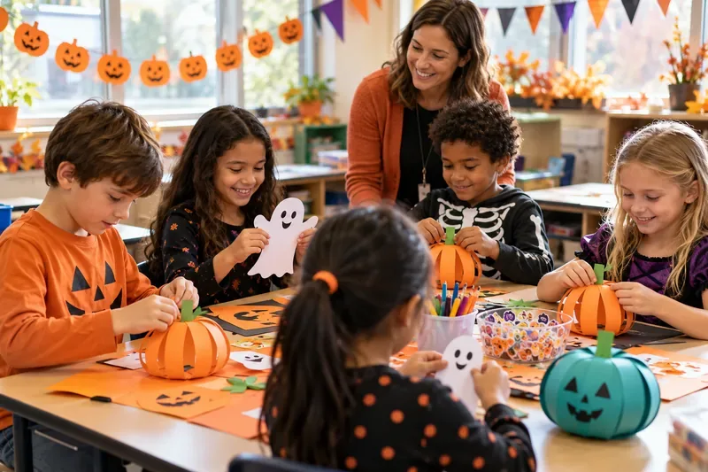
Plan a fun elementary school Halloween party with easy crafts, games, inclusive treats, costume tips, and a sample 75-minute classroom schedule.
Compare realistic side hustles for 2026 by startup cost, fees, flexibility, and income control, with current tax and scam guidance from official sources.
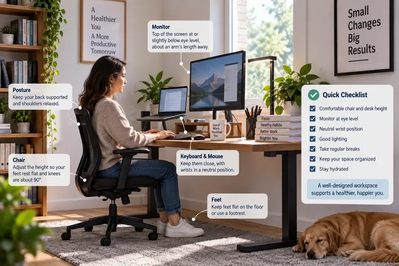
Upgrade your home office ergonomics with current OSHA and NIOSH guidance on chair fit, monitor height, laptop setup, lighting, and movement.